
Ultrasound Scans

Share images directly to your device
General diagnostic pelvic ultrasound (non-pregnant)
This scan can be performed at any point in the menstrual cycle, although scheduling in the early follicular phase (days 5–10) may improve visualization of the endometrium and ovaries. It is indicated for symptoms such as pelvic pain, abnormal bleeding, infertility, or suspected masses. The scan uses a transabdominal and/or transvaginal transducer to assess the uterus, endometrium, ovaries, adnexa, and pelvic spaces.
Pelvic ultrasound for deeply infiltrating endometriosis (non-pregnant)
Best performed in the early to mid-follicular phase of the cycle when hormonal changes are minimal. This scan is used to map suspected endometriosis, including ovarian endometriomas and deep pelvic disease involving bowel, bladder, or uterosacral ligaments. A high-resolution transvaginal scan is usually used, sometimes with bowel preparation, to identify abnormal nodules, adhesions, and organ involvement.
Dating / viability ultrasound
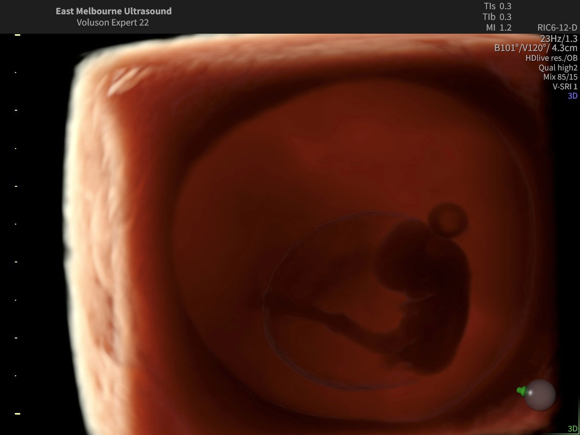
Performed in early pregnancy, ideally between 6–10 weeks’ gestation. The purpose is to confirm the presence of an intrauterine pregnancy, establish gestational age using crown-rump length (CRL), and assess fetal viability by demonstrating cardiac activity. The scan also excludes multiple pregnancy or early complications such as miscarriage or ectopic pregnancy.
Nuchal translucency ultrasound and early assessment of fetal anatomy
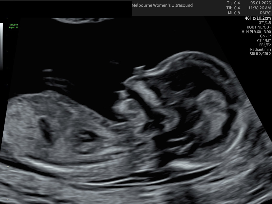
Conducted between 11+0 and 13+6 weeks’ gestation (CRL 45–84 mm). It measures the nuchal translucency and may assess nasal bone, ductus venosus, and tricuspid flow to estimate risk of chromosomal abnormalities. The scan also provides early evaluation of fetal anatomy, placental position, and chorionicity in multiple pregnancies.
Cervical surveillance ultrasound
Usually performed from 14–26 weeks’ gestation in women at increased risk of preterm birth (e.g., previous preterm birth, cervical surgery, multiple pregnancy). A transvaginal scan is used to measure cervical length and assess funnelling. It helps identify women who may benefit from interventions such as cerclage, progesterone, or pessary.
Early morphology ultrasound
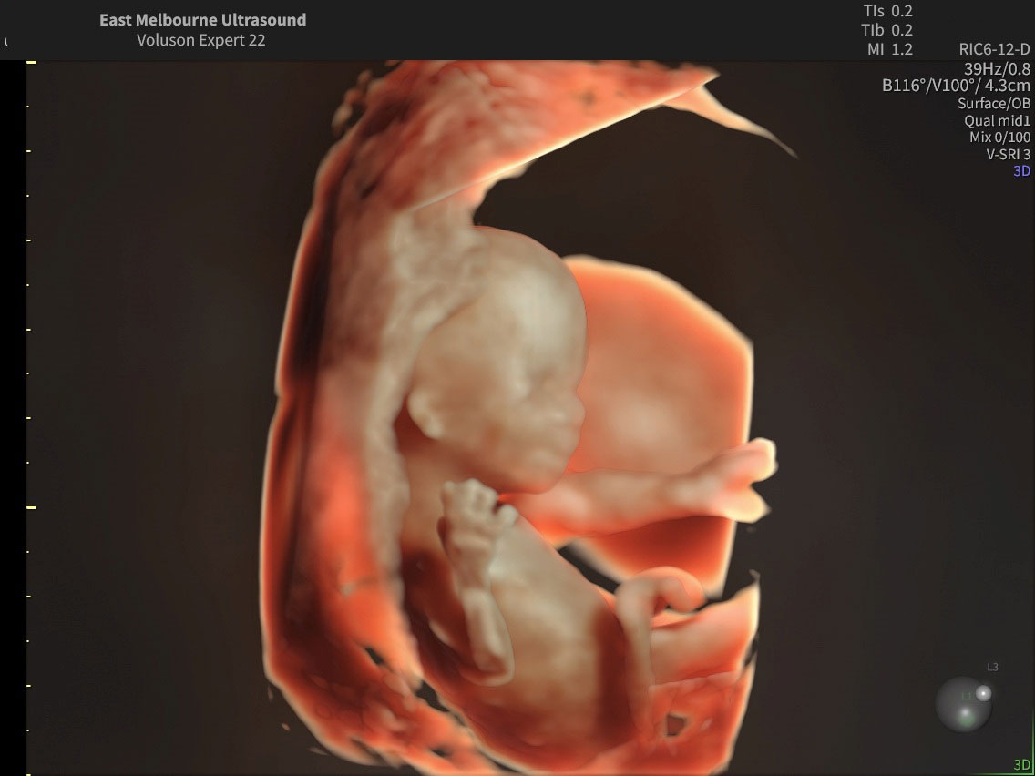
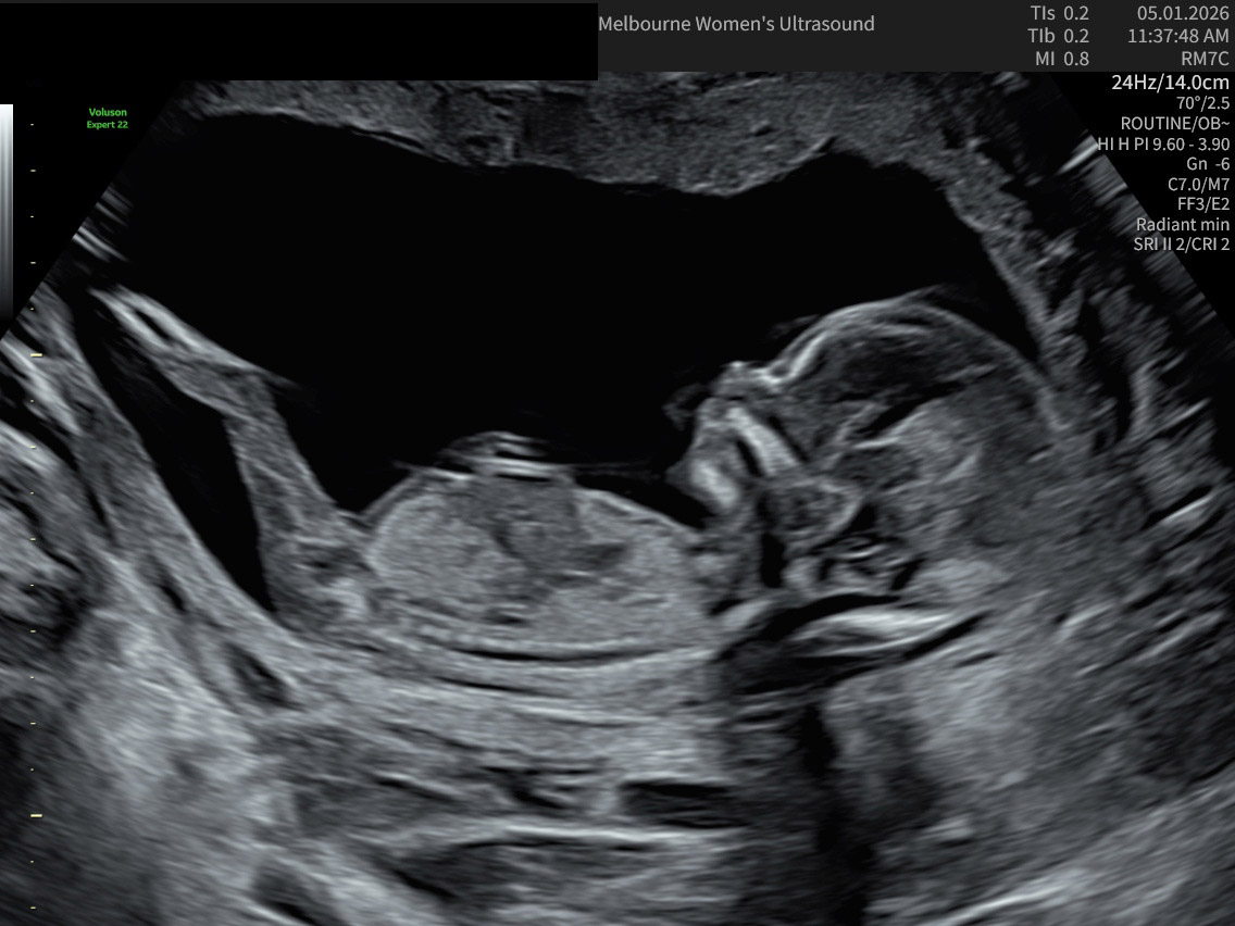
Performed at 14–16 weeks, often as an adjunct when early anomaly detection is indicated (e.g., high-risk history, previous abnormal findings). It evaluates major structural anomalies that may already be detectable, such as neural tube defects or abdominal wall defects. The scan also reassesses growth, amniotic fluid, and placenta.
Routine midtrimester fetal morphology ultrasound
The standard mid-trimester morphology scan is performed at 18–22 weeks, ideally around 20 weeks. It provides a systematic assessment of fetal anatomy, growth, placental location, amniotic fluid, and cervical length. This scan is the main screening for congenital anomalies and placental abnormalities.
Fetal growth and wellbeing ultrasound
Usually performed in the third trimester (from 28 weeks onwards, often at 32–36 weeks, or earlier if clinically indicated). The scan measures biometry to calculate estimated fetal weight and assesses growth trends. Wellbeing parameters include amniotic fluid, Doppler studies (umbilical artery, middle cerebral artery), fetal movements, and placental function.
Multiple pregnancy ultrasound
These scans are performed throughout pregnancy, with timing depending on chorionicity: dichorionic twins every 4 weeks from 20 weeks, monochorionic twins every 2 weeks from 16 weeks. They assess growth of each fetus, chorionicity, amnionicity, amniotic fluid, cord insertions, and placental location. The scans aim to detect complications unique to multiple pregnancies, such as twin-to-twin transfusion syndrome.
Chorionic villus sampling (CVS)
Performed between 11- and 14-weeks’ gestation. Ultrasound guidance is used to insert a needle or catheter through the abdomen or cervix to obtain placental tissue. The test provides early genetic diagnosis for chromosomal abnormalities or single-gene disorders, and ultrasound ensures safety by guiding the procedure and monitoring the fetus.
Amniocentesis
Usually performed from 15 weeks onwards. Under continuous ultrasound guidance, a fine needle is inserted through the maternal abdomen into the amniotic sac to withdraw fluid. This procedure allows genetic, biochemical, and infectious disease testing, and ultrasound ensures correct needle placement while avoiding harm to the fetus and placenta.
